Swift Ninja Catheter: Does what it says on the tin

65 year old with a left femoral renal metastasis lesion

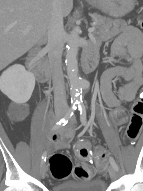
Angiograms showing vascularity of the lesion. The two annotated images show the tortuosity of the vessel supplying the lesion. The dotted orange coloured line shows the path of the vessel. It was not possible to cannulate this artery with conventional microcatheters.

Adequate catheter position attained using the Swift Ninja Catheter by Merit Medical. Final image showing reduced vascularity.

Saccular aortic aneurysm

60 year old male patient presented to the hospital with severe pain in the adbomen.

He underwent a CT scan which showed a 5 cm dilatation of his large artery in the abdomen called aorta (saccular infrarenal aortic aneurysm). 

This was treated by interventional radiology using pinhole techniques. A small access was placed in the right groin artery (right CFA) and repair was done endovascularly using a Medtronic Renu cuff. The device was placed percutaneously and the access site closed using Proglide closure device by Abbott.

Whole procedure done under local anaesthetic and completed in 20 minutes. Patient was asymptomatic following the procedure and discharged the following day. 

Patient with haemoptysis

Patient coughing up large amount of blood

66 year patient with cavitating left lung lesion likely mycetoma and haemoptysis.

CT Scan

Angiogram showing abnormal vessels


CT scan showed a cavitating lesion in the left upper lobe with a filling defect in the middle. Appearances suggestive of an aspergilloma. Patient became unstable due to large volume of haemoptysis.

Selective angiogram of the left bronchial artery shows the abnormal arteries supplying the abnormal area .

Embolisation performed using 500 to 700 micron PVA particles. No filling of the abnormal vessels following the procedure with immediate relief in haemoptysis.

Angioplasty

Thrombolysis of bypass graft

Elderly patient with previous fem distal bypass.

Angioplasty

Initially presented with a stenosis at proximal anastomosis. This was treated with angioplasty.


angiogram

Thrombolysis and Stent
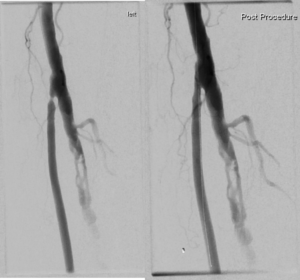
Two weeks later represented acutely with thrombosed graft. Due to underlying problems with the graft decision made to attempt to re-canalise the native arteries.

Subintimal angioplasty of native SFA and popliteal arteries done.

Zilver PTX stent placed in the SFA and a Supera stent in the popliteal artery. Single vessel inline flow to posterior tibial artery. Chemical thrombolysis with Alteplase to clear the residual thrombus in the posterior tibial.


Triphasic flow is seen on Doppler three months later.

CT Angiogram

Subclavian Artery Stent

Acute Cold Left Hand

48 yr old smoker presented with a cold left arm.

CT Angiogram
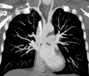
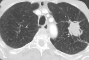
CT shows likely embolic disease in the left subclavian artery. There is high risk of further distal embolisation to hand or cranial embolisation via the left vertebral artery.


A ‘lifestream’ balloon expandable covered stent placed from left brachial artery approach with excellent outcome. Patients hand felt warm immediately after the procedure. Access site closed using ‘Proglide’ and patient discharged the following day.